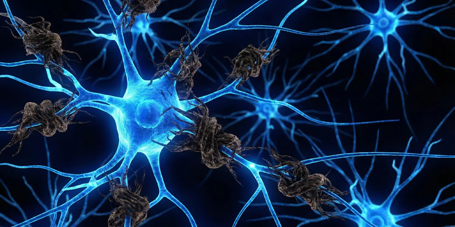
Brain scans reveal tau buildup in nearly two-thirds of late-onset psychosis patients
Background on Late-Onset Psychosis
Psychotic symptoms such as hallucinations and delusions usually appear in adolescence or young adulthood. However, in a subset of individuals, these signs emerge for the first time after age 40, sometimes after 60. These cases are termed late-onset psychosis. This form often differs from early-onset psychosis. Older patients tend to have more prominent persecutory delusions, such as believing others wish to harm them, while showing fewer negative symptoms like flat affect or social withdrawal, and less disorganized thinking. These differences led to the hypothesis that late-onset psychosis may involve brain changes distinct from those seen in early forms.
Wellness Alert
 Emotional Intelligence Linked to Workplace Resilience, New Study Finds
 Take Charge of Your Health: Why You Are Your Best Medical Advocate
 Anxiety in the practice room: How performance stress shapes student well-being
 Stop Believing Everything You Hear: The Hidden Cost of Unverified Information
Earlier small-scale brain imaging studies identified alterations in white matter, especially in posterior brain regions, and changes in blood flow in some individuals with late-onset psychosis. A large Swedish study showed that people developing psychosis after age 60 have an increased risk of dementia in subsequent years. Postmortem analyses of deceased patients revealed multiple neurodegenerative conditions in late-onset psychosis cases, including tauopathies distinct from typical Alzheimer’s pathology. Most of this evidence comes from autopsies, as until recently, tools for detecting the full spectrum of tau buildup in living people were limited.
Prior Imaging and Neuropathology Findings
The research team had previously developed a radioactive tracer called florzolotau, used in positron emission tomography (PET) imaging, which binds to tau protein and can identify both Alzheimer’s-associated tau aggregates and varieties seen in other neurodegenerative diseases. The study was conducted by researchers Manabu Kubota and Shin Kurose from Japan’s National Institute for Quantum Science and Technology, together with colleagues. They hypothesized that amyloid-beta and tau buildup would be more frequent in late-onset psychosis patients compared to healthy adults of the same age.
The study included 37 patients diagnosed with late-onset psychosis and 47 healthy adults as controls. The average age of patients was 70 years, while the healthy group averaged 66 years. Of the total patients, 27 were women, and in the control group, 22 participants were female. Most patients were diagnosed with schizophrenia (20 cases) or delusional disorder (13 cases), and none showed signs of cognitive impairment or neurological disease at symptom onset.
In 24 cases, symptoms began after age 60; in the remaining 13, onset occurred between ages 40 and 60. Compared to the healthy group, patients scored lower on cognitive function tests and had lower educational levels.
Study Design and Methods
All participants underwent magnetic resonance imaging (MRI) and two PET scans using small amounts of radioactive substances to bind to specific brain proteins. One scan targeted amyloid-beta detection, while the other used the florzolotau tracer to identify tau presence. Although both proteins exist naturally in the brain, abnormal accumulations can lead to plaque formation in amyloid’s case—characteristic of Alzheimer’s—and tangle formation in tau’s case, structures seen not only in Alzheimer’s but also in many other neurodegenerative diseases.
Three raters analyzed amyloid-beta PET images and classified each participant as positive or negative for this protein using standardized procedures from the Japanese Neuroimaging Initiative for Alzheimer’s Disease. For tau positivity, a voxel-by-voxel computerized analysis of each PET image was used.
The results showed that 13 of 37 patients (35%) were classified as amyloid-positive, while only one participant in the healthy group of 47 (2%) showed this trait. All amyloid-positive patients developed psychosis symptoms after age 60. Tau accumulation proved even more widespread: 65% of patients (24 of 37) were tau-positive, compared to just 15% of healthy participants (7 of 47).
Results
Patients were divided into three roughly equal groups by number. Twelve of the 13 amyloid-positive patients were also tau-positive, a combination consistent with Alzheimer’s pathology. Half of the 24 amyloid-negative patients showed tau buildup without amyloid, a feature that may indicate non-Alzheimer’s tauopathies. The remaining 12 patients showed neither amyloid nor tau accumulation.
Tau distribution patterns varied considerably from patient to patient.
More stories: